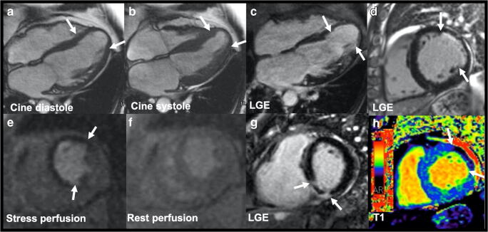
Fig. 1.
Ischemic cardiomyopathy images in cine, late gadolinium enhancement (LGE), stress perfusion, microvascular obstruction, and hemorrhage. a, b Still frames from 4-chamber cine in end-diastole and end-systole showing hypokinesis of the mid-ventricle to the apex with an apical aneurysm (between arrows) in a 65-year-old woman. c, d LGE images showing subendocardial delayed enhancement in the apical walls (between arrows) of the 65-year-old woman and subendocardial LGE in a 78-year-old man with an old myocardial infarction (MI). e, f Stress perfusion image showing perfusion defects of the anterior wall and the septum compared to the rest perfusion image, suggesting reversible ischemia (between arrows) in the 65-year-old woman. g LGE image showing microvascular obstruction in the basal inferoseptum (between arrows) in a 40-year-old man with acute MI. h T1 mapping showing the dark core of intramyocardial hemorrhage (between arrows) in the 78-year-old man with the corresponding LGE slice in image (d)

