Fig. 2.
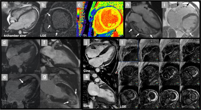
Non-ischemic cardiomyopathy. Dilated cardiomyopathy images in cine, late gadolinium enhancement (LGE), and T1 mapping. a CMR end-diastolic frames from 4-chamber cine showing biventricular dilation in a 23-year-old man with chronic heart failure. b LGE imaging showing mid-myocardial delayed enhancement in the septum and the inferior wall (arrows). c T1 mapping shows diffusely elevated signals consistent with diffuse fibrosis. Hypertrophic cardiomyopathy (HCM) presentations in cine and late gadolinium enhancement. d, f 4-chamber or 2-chamber long-axis cine images showing different phenotypes of HCM. e, g LGE images showing patchy delayed enhancement (arrows) in corresponding patient cine views. d, f Asymmetric septal hypertrophy. e, g Apical HCM. Arrhythmogenic right ventricular cardiomyopathy. h Still frame from 4-chamber cine in end-diastole showing dilated, trabeculated right ventricle with regional thinning and microaneurysm (white arrows) in a 76-year-old man. i Late gadolinium enhancement image showing enhancement in the right ventricular wall (black arrows). The left ventricular LGE was due to a left circumflex occlusion in this patient, not left ventricular involvement in ARVC (white arrow). Amyloidosis. j, k Still frames from 2-chamber and 4-chamber cines showing bi-atrial enlargement, increased wall thickness of left ventricle, and trace pericardial effusion in a 65-year-old man. l Serial short-axis late gadolinium enhancement images showing diffuse transmural enhancement in the basal to mid-left ventricular myocardium, gradually decreasing to a global subendocardial pattern in the mid-ventricle. Note that the blood pool is dark which is related to abnormal blood pool gadolinium dynamics in the presence of diffuse amyloid deposition in the body

