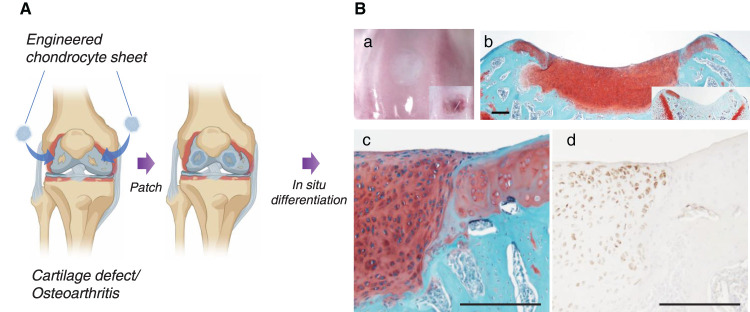
Figure 2. Cartilage regeneration with engineered human juvenile polydactyly cartilage-derived chondrocyte sheets.
(A) Schematic images of chondrocyte sheet transplantation to knee defects. (B) Repaired trochlear groove cartilage with human juvenile polydactyly cartilage-derived chondrocyte sheet. In situ hyaline cartilage maturation occurs within 4 weeks in rodent focal cartilage defect models. (a) Macroscopic image (right corner box shows defect only control). (b) Safranin O staining of nude rat trochlear groove (right corner box shows defect only control with fibrotic tissue formation). (c) Magnified image of Safranin O staining at regenerated cartilage and host tissue interface. Note no gap is observable at the interface. (d) Human vimentin antigen-specific immunostaining, suggesting that regenerated cartilage originates from transplanted human cells. Bars: 200 μm.

