
Figure 4.
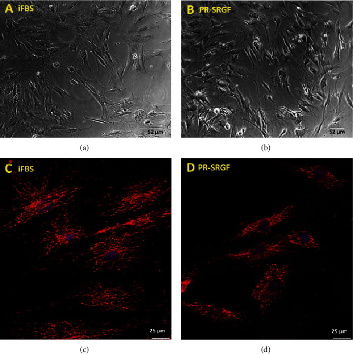
(a, b) Light microscopy of living unstained MSCs (10x lens) cultivated for 48 h in medium supplemented with 10% heat inactivated FBS (iFBS) (a) and 7.5% PR-SFGF (b). (c, d) Confocal images of mitochondrial network of fixed MSC (63x lens) cultivated for 48 h in medium supplemented with 10% heat inactivated FBS (iFBS) (c) and 7.5% PR-SFGF (d).