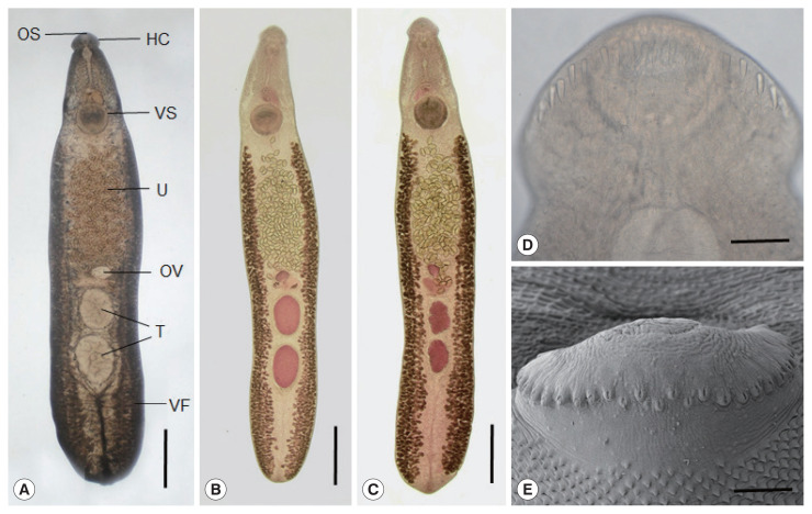
Fig. 2.
Echinostoma mekongi adult worms obtained from an experimental hamster at day 20 post-infection. (A) An adult fluke showing the head collar (HC), oral (OS) and ventral suckers (VS), uterus (U) filled with eggs, ovary (OV), globular testes (T), and vitelline follicles (VF). Ventral view. Unstained. Scale bar=0.1 mm. (B, C) Other worms showing the same organs in Fig. 2A. Note the 2 globular (B) or slightly lobulated testes (C) and non-confluent vitellaria beyond the testicular field (B, C). Ventral view. Stained with Semichon’s acetocarmine followed by clearing in glycerin-alcohol and mounting in glycerin-jelly. Scale bar=0.1 mm (B, C). (D, E) Light microscopic (D) and scanning electron microscopic (E) views of the head collar with collar spines. Note the 2 alternating rows of the dorsal collar spines. Dorsal views. Scale bars=100 μm (D, E).

