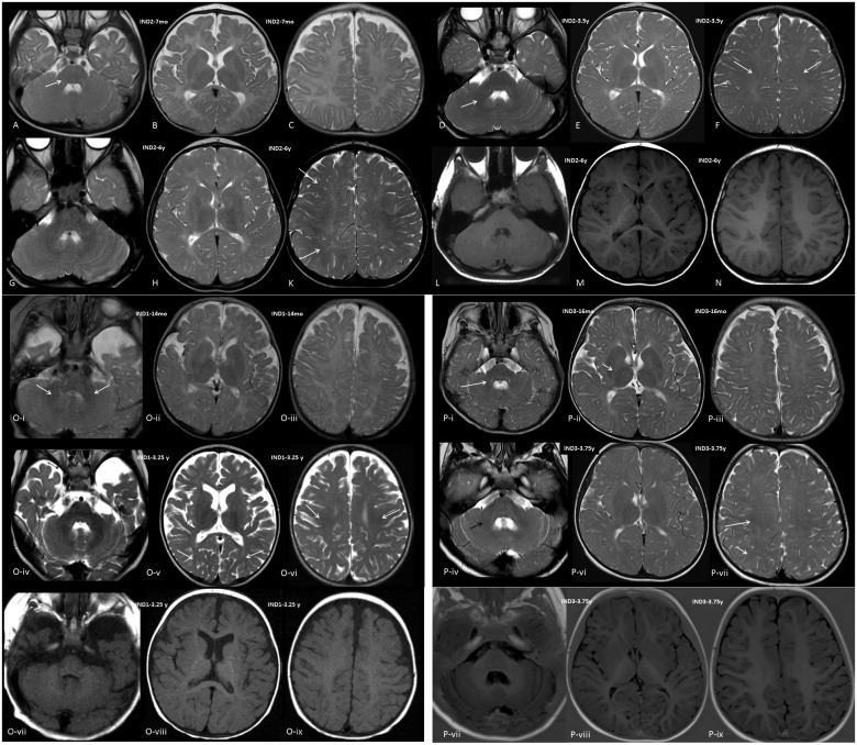
Figure 1.
Cranial MRI images. Images shown are of Individual 2 (A–N), Individual 1 (O) and Individual 3 (P). (A–C) T2-weighted axial images of Individual 2 at the age of 7 months, (D–F) T2-weighted axial images at age 3.5 years, (G–K) T2-weighted axial images, at age 6 years, and (L–N) T1-weighted axial images at age 6 years at the level of the cerebellum (A, D, G and L), diencephalon (B, E, H and M) and centrum semiovale (C, F, K and N). There is no myelin equivalent at 7 months on T2-weighted images except for a mildly lower signal in the cerebellar peduncles (arrow); at 3.5 years, central white matter shows some lower signal (arrows), as well as the central cerebellar white matter (short arrow); at 6 years, lower signal is seen in the more peripheral frontal and occipital white matter (arrows); T1-weighted images at 6 years appear near normal, U-fibres are not clearly myelinated, however, and signal is slightly less hyperintense than normal. [O(i–iii)] T2-weighted axial images of Individual 1 at the age of 14 months, (iv–vi) T2-weighted axial images at age 3.25 years, (vii–ix) T1-weighted images at age 3.25 years at the level of the cerebellum (i, iv and vii), diencephalon (ii, v and viii) and centrum semiovale (iii, vi and ix). There is no myelin equivalent at 14 months on T2-weighted images except for a lower signal in the cerebellar peduncles (arrows) and cerebellar white matter, central white matter may show some minimally lower signal; at 3.25 years, central white matter shows lower signal (arrows), as well as the more peripheral occipital white matter (short arrows), the cerebellar white matter is clearly of low signal. T1-weighted images at 3.25 years show hyperintensity of white matter with signal; however, less hyperintense than normal; U-fibres are not clearly myelinated. There is evidence of some global atrophy of the telencephalon (somewhat reduced white matter volume and enlarged sulci). [P(i–iii)] T2-weighted axial images of Individual 3 at the age of 16 months, (iv–vi) T2-weighted axial images and (vii–ix) Inversion recovery axial at age 3.75 years, at the level of the cerebellum (i, iv and vii), diencephalon (ii, v and viii) and centrum semiovale (iii, vi and ix). There is no myelin equivalent at 16 months on T2-weighted except for a lower signal in the cerebellar peduncles and cerebellar white matter (arrow), and minimally in the posterior limb of the internal capsule (PLIC; short arrow); at 3.75 years, central white matter shows lower signal (arrow), as does the PLIC and the more peripheral occipital white matter (short arrow), the cerebellar white matter is clearly of low signal (black arrow); inversion recovery images at 3.75 years appear near normal, U-fibres are not clearly myelinated; however, as evident through the somewhat blurry differentiation between cortex and white matter; and signal is somewhat less hyperintense than normal. IND = individual; mo = months; y = years.

