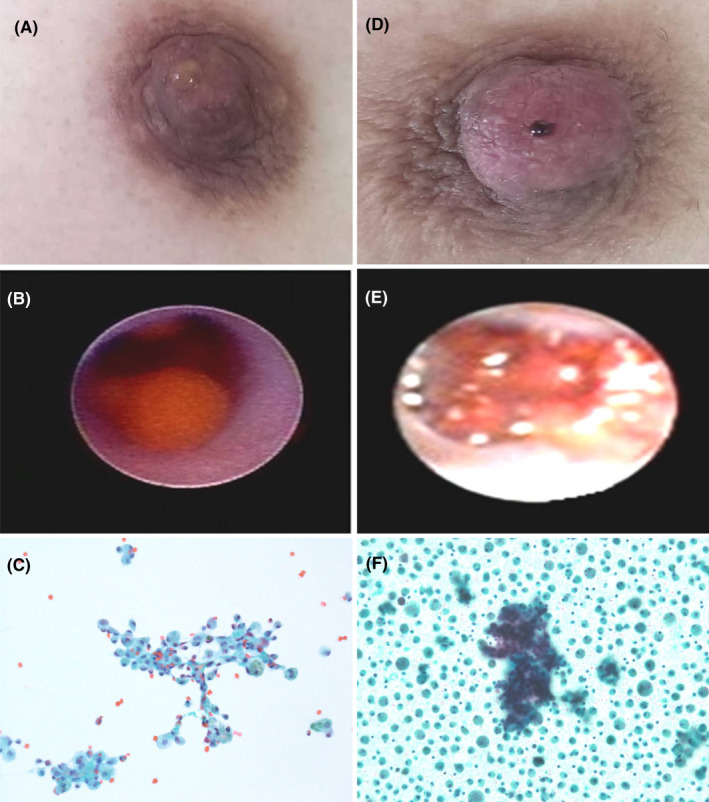
FIGURE 1.

Representative examples of benign (A–C) and malignant (D–F) nipple discharge. (A) PND with light yellow serous liquid; (B) Fiberoptic ductoscopy system showed inductal pappiloma; (C) TCT showed benign ductal cells and foamy macrophages in background of blood (Papanicolaou stain, ×200); (D) PND with spontaneous bloody fluid; (E) Fiberoptic ductoscopy system showed inductal carcinoma; (F) TCT showed focally dyshesive three‐dimensional cell groups of large pleomorphic cells in background of foamy macrophages (Papanicolaou stain, ×200)
