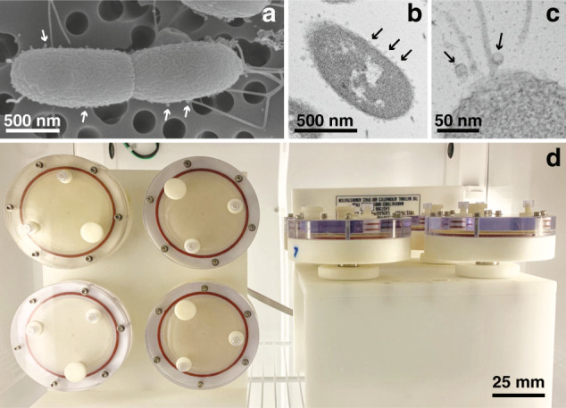
Fig. 1. Overview of the beneficial microbe Vibrio fischeri morphology and experimental conditions.

a Scanning electron micrograph of the wild-type V. fischeri depicting the presence of outer-membrane vesicles (OMVs) on the cell surface (arrows). b Transmission electron micrograph (TEM) of V. fischeri during exponential growth producing numerous OMVs during exponential-phase growth. c Higher magnification TEM visualizing OMVs associated with the bacteria flagella. d Rotary cell culture system with high-aspect ratio vessels containing V. fischeri cultures in the modeled microgravity (left) and gravity (right) control positions.
