Figure 1.
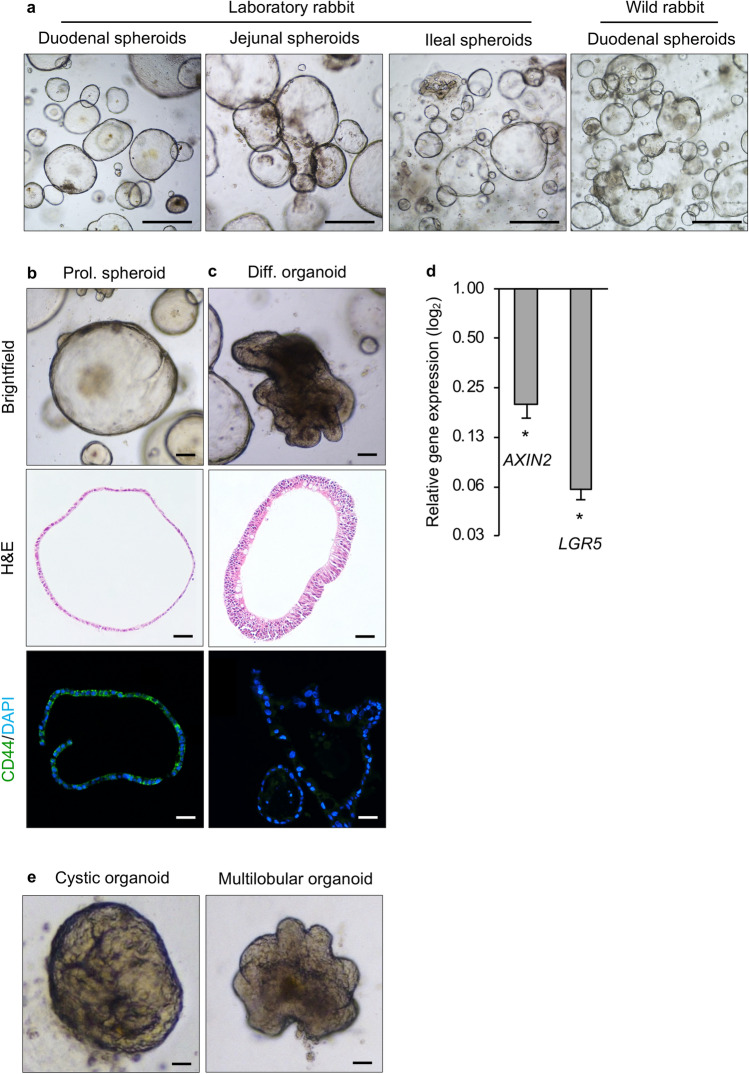
Morphology and characteristics of rabbit small intestinal organoids. (a) L-WRN-conditioned medium supported the growth of spheroids from duodenum, jejunum and ileum spheroids from a laboratory rabbit, and duodenum spheroids from a wild rabbit. Scale bars = 500 μm. (b) Proliferating rabbit duodenal spheroids and (c) organoids were imaged either unstained (brightfield), after staining with hematoxylin and eosin (H&E) or after immunostaining with a CD44-specific (green) antibody; nuclei were counterstained with DAPI (blue). (d) Expression of stem cell-related genes (AXIN2 and LGR5) in differentiated organoids. Data are presented as fold change (2−ΔΔCt) in gene expression from undifferentiated spheroids, calculated from three individual cell culture wells with three technical RT-qPCR replicates each. Error bars represent standard errors of the mean. Student’s t-test was performed to assess the statistical significance; only statistically significant differences are shown (*p < 0.05). (e) Representative images of organoids after four days of culturing in differentiation medium; the images show representative organoids with either a cystic (left panel) or multilobular morphology (right panel). Scale bars = 100 μm. b–e Used duodenal spheroids/organoids from a single laboratory rabbit.

