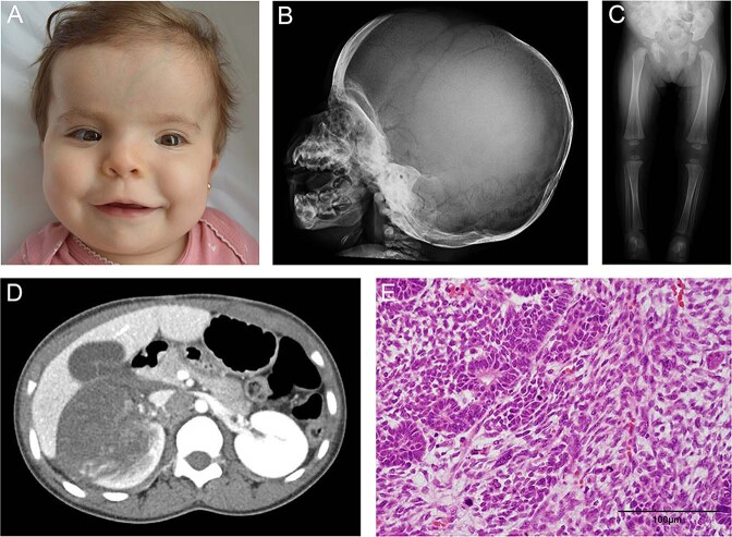
Fig. 1. Clinical, radiographical and histological data from cases 2 and 3.
a Photograph of case 3 showing typical features of OSCS including macrocephaly, frontal bossing, hypertelorism, and a long philtrum. b Macrocephaly and cranial sclerosis on skull X-ray in case 3. c Metaphyseal striations in the long bones of the lower limbs on X-ray from case 3. d Abdominal computerized tomography scan obtained at diagnosis from case 2 showing a large right-sided renal mass, perinephric hematoma, and retrocaval lymphadenopathy. e H&E-stained histology from case 2 of resected tumor showing Wilms tumor with diffuse anaplasia.

