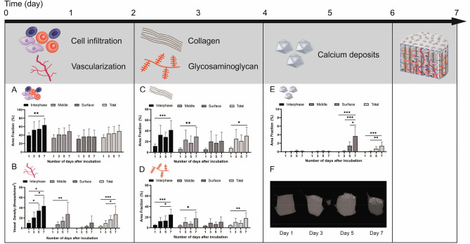
Figure 2.
Time course regarding the occurrence and impact of key factors on the calcification process analyzed in collagen scaffold. The timing of the various factors involved was investigated (A–F) using the CAM assay. A marked increase in cell infiltration and vessel density in the scaffold was observed on day 1 (A, B). On day 3, there was a distinct increase in collagen and glycosaminoglycan (GAG) content (C, D) and from day 5 on, an accumulation of calcium deposits could be sufficiently detected (E), which could also be shown by microCT analysis (F). Schematic scaffold with the localization of key factors is shown after 7 days of incubation on the CAM.

