Figure 6.
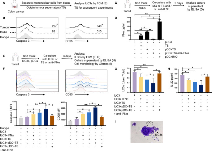
pDCs can affect the survival of ILC3s in the tumor-like microenvironment. (A, B) MNCs from tumor and distal regions from three patients with colon cancer were prepared. Flow cytometric analysis was performed for the expression of apoptosis-related genes caspase 3 and CD95 on ILC3s in the tumor and distal tissue control groups. (C, D) pDCs from normal tonsil tissue were prepared. TS was added to the pDC culture system and the culture supernatant was collected to detect the secretion of cytokine IFN-a from pDCs by ELISA; IMQ was used as a positive control; anti-IFNα was used to neutralize IFN-a. TS is from tumors of three patients with colon cancer. (E–I) ILC3s and pDCs from normal tonsil tissue were prepared. TS, IFN-a, and anti-IFNα was added to the culture system of ILC3s and pDCs. Flow cytometric analysis of the expression of apoptosis-related genes (F) and the survival rate of ILC3s (G) are shown. ELISA was used to detect the secretion of IL-22 in co-culture supernatant (H). Giemsa staining was used to detect the morphology of ILC3s and pDCs (I) (magnification, 50×). Each experiment was repeated three times. A paired t-test and Spearman test were used for statistical comparisons. *P < 0.05; **P < 0.01.

