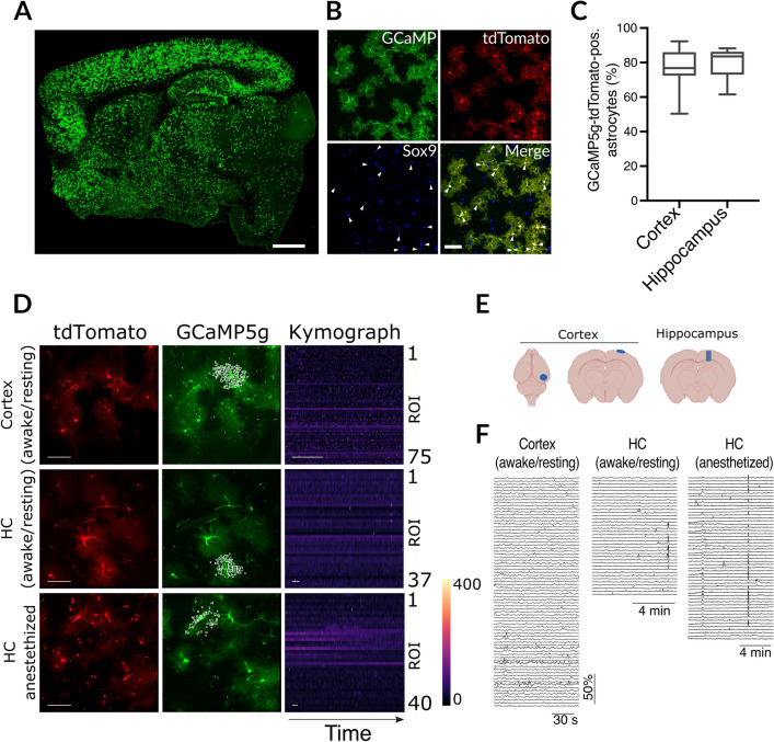
Figure 3.
Calcium imaging. (A,B) Fluorescent imaging of Slc1a3-CreERT2::GCaMP5g-tdTomato-loxP using antibodies against GFP, the astroglial marker Sox9, and the native fluorescence of tdTomato, showed widespread and strong Cre recombination specifically in astrocytes (arrowheads; scale bars, 1 mm and 50 µm, respectively; Sox9 nuclear marker was used to identify astrocytes. (C) Quantification of recombination efficacy in hippocampus and cortex; reporter expression was normalized to all Sox9-positive cells. (D) Representative examples of calcium activity under awake/resting conditions in the cortex, and under awake/resting conditions or anesthesia in the hippocampus (HC). No changes were seen in tdTomato native fluorescence (scale bars, 50 µm), indicating stable imaging conditions over time, while activity-evoked calcium changes could be detected using GCaMP5g native fluorescence. The kymographs represent color-coded calcium changes over time (scale bars, 30 s) in distinct regions of interest (ROI) from individual astrocytes (marked by white boxes in the GCaMP5g images). (E) Locations of cranial windows implanted over cortex and hippocampus. (F) Individual traces for all conditions and regions. The traces correspond to ROIs and kymographs depicted in D. Mice were 4 months of age.

