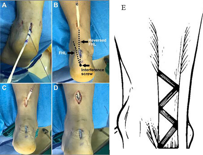
Figure 3.
(A) The flexor hallucis longus (FHL) tendon is pulled into the midline incision using a PDS-2 suture loop (Depuy Ethicon). (B) The reversed FHL tendon is passed through the tunnel to the distal end of the proximal incision. The left dotted line indicates the original FHL tendon, and the right dotted line indicates the reversed FHL tendon. (C, D) The FHL tendon is woven through the proximal stump for augmentation. (E) The FHL tendon is woven into the proximal stump in a Z-shaped tunnel.

