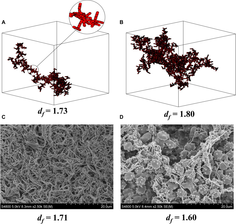
FIGURE 1.
df and clot microstructure. Computer modeling of fractal structures of blood clots at the gel point from (A) healthy individuals and (B) individuals with vascular inflammatory disease. Electron microscopy images of fractal dimension (df) and clot microstructure in whole blood (C) pre and (D) post 1 wk of oral dual antiplatelet therapy (75 mg Aspirin and 10 mg Prasugrel) in healthy individuals. The pictures (C,D) clearly show how inhibition of platelet activity alters clot microstructure and mass as indicated by df. Note that a small change in df results in a large increase in mass at the gel point for the developing clot. (A,B) are reproduced from Curtis et al. (2011).

