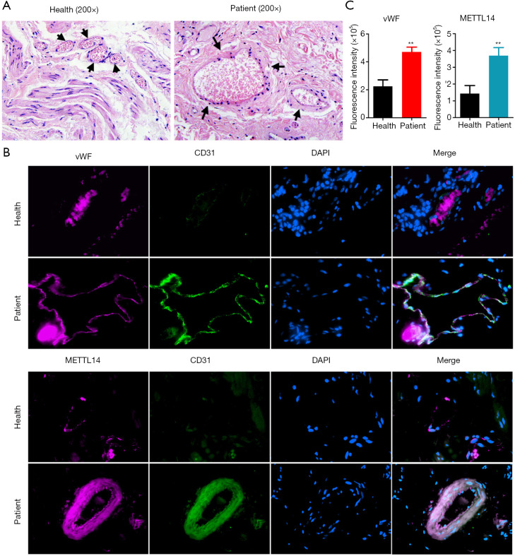
Figure 3.
Vascular markers and METTL14 were highly expressed in hemorrhoids. Pathological examination results using (A) hematoxylin-eosin (HE) staining. The arrows indicate blood vessels. Immunofluorescence staining results in each group (B). Magnification: 200×. The proportion of vWF+ and CD31+/METTL14+ cells in (C) hemorrhoids was significantly higher than that in normal tissues. **P<0.01 vs. healthy tissue; t-test; n=4.

