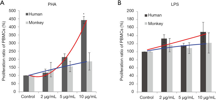
Figure 1.
Lymphocyte proliferation in response to PHA or LPS. Proliferation by human and cynomolgus PBMCs were measured via CCK-8 assay. An F-test of two regression line equations was used to compare trends for proliferation rates between human and monkey cells. Lines in red and blue indicate trend curves for proliferation of the human and monkey PBMCs, respectively. (A) The trend for the proliferation of PBMC was significantly different between humans and cynomolgus monkeys after PHA stimulation (*, P<0.05). (B) No significant difference in trends for proliferation was noted between human and cynomolgus monkey cells after LPS stimulation. PHA, phytohemagglutinin; LPS, lipopolysaccharide; PBMC, peripheral blood mononuclear cells; CCK-8, Cell Counting Kit-8.

