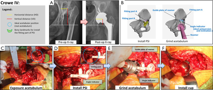
Figure 3.
Details of application of the new PSI for patients with Crowe IV developmental dysplasia of the hip (DDH). (A) Preoperative and postoperative pelvic radiographs of a typical case. The centre of rotation (COR), horizontal distance (HD) and vertical distance (VD) have returned to normal after surgery. (B) Schematic diagram of the two main steps (installation of PSI and grinding down of the acetabulum) of the new PSI surgical method. (C,D,E,F) Application of the PSI during surgery.

