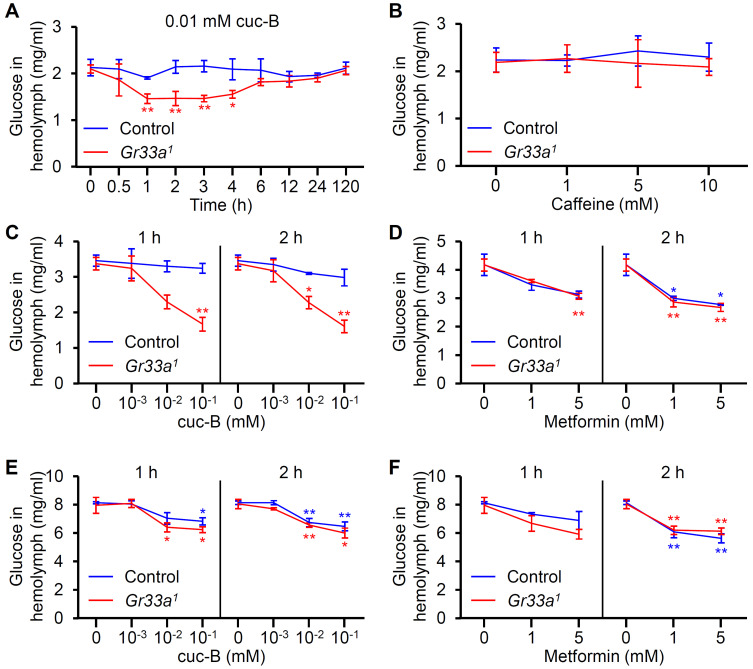
Fig. 2. Effect of cuc-B, caffeine, and metformin treatment on hemolymph glucose levels.
(A) Hemolymph glucose levels in control and Gr33a 1 males after being fed with standard cornmeal mixed with 0.01 mM cuc-B (n = 4). (B) Hemolymph glucose levels in control and Gr33a 1 male flies after 1 h of being fed with standard cornmeal mixed with the indicated caffeine concentrations (n = 4). (C) Hemolymph glucose levels in control and Gr33a 1 flies 1 h and 2 h after being fed with 3% sucrose with the indicated cuc-B concentrations (n = 4). (D) Hemolymph glucose levels in control and Gr33a 1 flies 1 h and 2 h after being fed with 3% sucrose with the indicated metformin concentrations (n = 4). (E) Hemolymph glucose levels in control and Gr33a 1 flies 1 h and 2 h after being fed with 20% sucrose with the indicated cuc-B concentrations (n = 4-5). (F) Hemolymph glucose levels in control and Gr33a 1 flies 1 h and 2 h after being fed with 20% sucrose with the indicated metformin concentrations (n = 4-5). All error bars indicate the SEM. (A and B) Asterisks indicate statistical significance (*P < 0.05, **P < 0.01), as determined by Student’s t-test comparisons with appropriate genetic controls. (C-F) Single-factor ANOVA coupled with Scheffe’s analysis was used as a post hoc test to compare multiple datasets (*P < 0.05, **P < 0.01). Each point was compared with 0 mM of each tested compound for each genotype.

