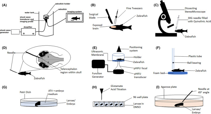
FIGURE 4.

Injury models utilized by the zebrafish TBI model. (A) Acoustic shock wave generated and bombarded onto zebrafish head, directed by B‐focal imaging system, (B) Mechanical lesion applied by surgically making a hole in the skull and using the fine tweezers to cut the specified brain region, (C) Injection of quinolinic acid into desired brain region under a dissecting stereomicroscope, (D) Telencephalon injury induced by inserting needle into the telencephalon region via nose, E) Pulsed high‐intensity focused ultrasound (pHIFU) generated and focally targeted toward zebrafish head through an ultrasonic membrane, (F) A steel ball‐bearing (weight) is dropped from a specified height through a plastic tube and unto the cranium of the zebrafish, (G) Larvae was incubated in a petri dish filled with atorvastatin (ATV) and embryo medium mixture, (H) Glutamate acid was titrated into wells of a 96‐well plate containing zebrafish larvae (each in one well), and (I) Stab lesion was inflicted in zebrafish larvae placed on agarose medium, via a needle angled at 45o toward the desired brain region
