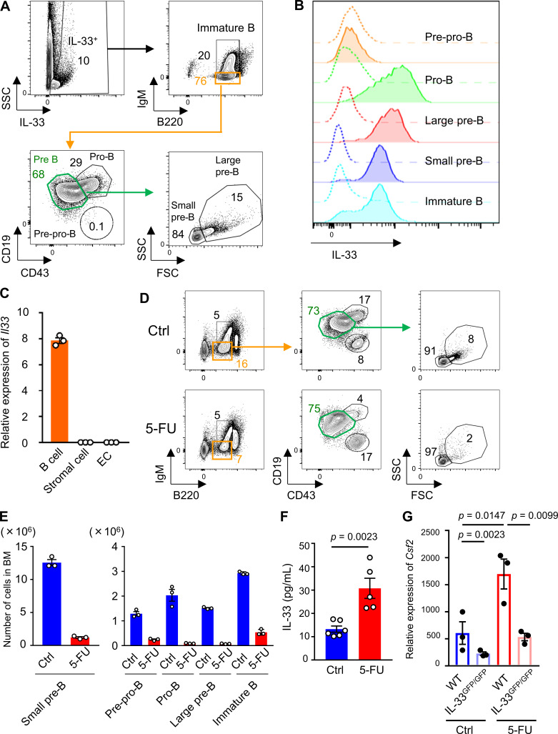
Figure 5.
IL-33 secreted by B cell progenitors activates BM ILC2s after 5-FU treatment. (A) Flow cytometry of BM cells from IL-33–GFP mice showing B cell progenitors, including pre-pro-B, pro-B, large pre-B, small pre-B, and immature B cells. Each number indicates the percentage of each fraction. Results shown are representative of three independent experiments. (B) IL-33–GFP levels in each B cell progenitor fraction of IL-33–GFP mice. Dashed lines represent background levels in WT mice. Results shown are representative of three independent experiments. (C) qRT-PCR analyses for the expression of Il33 in CD19+ B cells, stromal cells, and ECs sorted from WT mice; n = 3, representative of two independent experiments. (D) Flow cytometry plots of BM cells from control (Ctrl) or 5-FU–treated WT mice to evaluate the size of each B cell progenitor. Results shown are representative of three independent experiments. (E) The numbers of each B cell progenitor fraction from femurs and tibias of control or 5-FU–treated WT mice; n = 3, representative of two independent experiments. (F) IL-33 protein levels in femur analyzed by ELISA; n = 6 in control, n = 5 in 5-FU, representative of two independent experiments. (G) qRT-PCR analyses for the expression of Csf2 in BM ILC2s from WT and IL-33GFP/GFP mice; n = 3, representative of two independent experiments. In 5-FU–treated groups, mice were intravenously injected with 200 mg/kg 5-FU 2 d before euthanization. Results are shown as mean ± SEM. Statistical significance was determined by unpaired Student’s t test (F) or ANOVA with the Bonferroni post hoc test (G).

