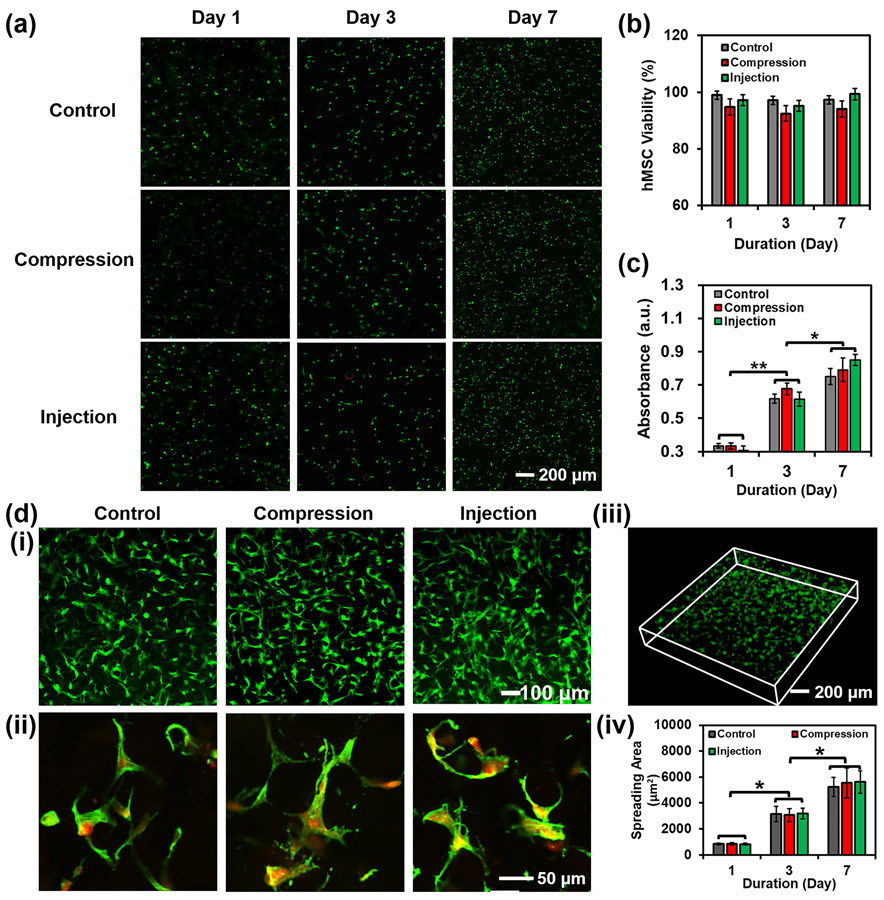
Figure 5.
Evaluation of hMSC viability and proliferation in the hydrogel constructs after compression and injection. (a) Fluorescence micrographs, (b) viability, and (c) proliferation of cells within the 3D-bioprinted porous hydrogel constructs before and after compression. Live cells were stained in green, while dead cells in red. The control group indicates the hydrogel constructs without compression and injection. (d) Fluorescence micrographs of cell spreading within the porous hydrogel constructs at (i) low and (ii) high magnifications. The cells were stained for F-actin (green) and nuclei (red). (iii) 3D reconstruction of spreading cells within a porous hydrogel construct. (iv) Calculated cell spreading areas on Day 1, Day 3, and Day 7 (n=3, *p<0.05).

