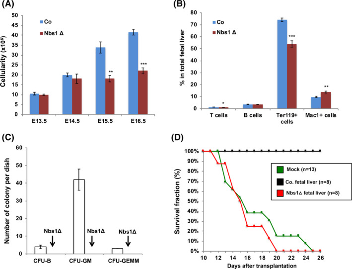
Figure 3.

Defective foetal liver haematopoiesis after Nbs1 deletion. A, Cellularity of foetal livers from control and Nbs1‐HSCΔ embryos at different developmental stages (n ≥ 3 for each group). B, Frequencies of T cells (CD3+), B cell (B220+), red blood cells (Ter119+), granulocytes and macrophages (Mac1+) in E15.5 control (Co) and Nbs1‐HSCΔ (Nbs1Δ) foetal livers (N = 3 for each genotype). C, Colony formation assay of control and Nbs1‐HSCΔ foetal liver haematopoietic cells. Please note that Nbs1‐HSCΔ foetal liver haematopoietic cells fail to form any haematopoietic colony in vitro. D, Kaplan‐Meier survival curve of lethally irradiated Ly5.1 mice transplanted with control or Nbs1‐HSCΔ foetal liver cells. Mock reconstitution (+PBS) is used to determine the lethal dose. Please note that Nbs1‐HSCΔ foetal liver haematopoietic cells could not reconstitute the haematopoietic system in lethally irradiated Ly5.1 mice. Note: *, P < .05; **, P < .01; ***, P < .001. Unpaired Student's t test is used
