Figure 4.

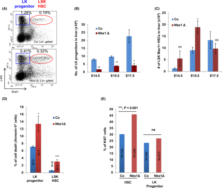
Cell fates of foetal liver HSCs and LK progenitors upon Nbs1 deletion. A, Representative FACS profile of HSCs and LK progenitors from E15.5 embryonic foetal livers of control (Co) and Nbs1‐HSCΔ (Nbs1Δ) mouse embryos. Frequencies of LK progenitors and LSK HSCs (in gated foetal liver cells) are shown. B, Absolute number of LK progenitors in foetal livers at different stage (N ≥ 3 for each group). C, Absolute number of HSCs in foetal livers at different stage (N ≥ 3 for each group). D, Apoptosis (Annexin V+) of LK progenitors and HSCs from E15.5 control and Nbs1‐HSCΔ foetal livers. ‘N’ denotes the number of embryos analysed. E, Ki67‐ cells in HSCs and LK progenitors at E16.5. HSCs and LK progenitors sorted from 2 controls and 2 mutant embryos are used for the analysis. N denotes the number of cells used for the quantification (chi‐square test is used. ***, P < .001; ns, not significant). Note: *, P < .05; **, P < .01; ***, P < .001. Unpaired Student's t test is used
