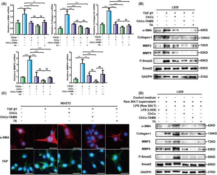
FIGURE 5.

The anti‐fibrogenic effects of the ChCo‐based scaffolds in vitro (A, B) L929 cells were precultured with the ChCo‐based scaffolds or TA for 48 h before induced with 10 ng/ml of TGFβ1 for 24 h, followed by mRNA detection of α‐SMA, collagen‐I, collagen‐III, MMP2, MMP9 or protein detection of α‐SMA, collagen‐I, MMP2, MMP9, p‐Smad2 and Smad2. (C) NIH3T3 cells were precultured with the ChCo‐based scaffolds or TA for 48 h before induced with 10 ng/ml of TGFβ1 for 24 h, followed by immunostaining of α‐SMA and FAP; scale bar, 20 μm. (D) The culture medium of RAW264.7 cells induced by 1 μg/ml LPS in the presence of the ChCo‐based scaffolds or TA for 24 h was collected and applied for culturing of L929 cells at 24 h. Immunoblotting was conducted to assess the variations of α‐SMA, collagen‐I, MMP2, MMP9, p‐Smad2 and Smad2. The representative statistical results were performed using one‐way ANOVA test and shown as means ± SEM from 3 independent experiments. *P < 0.05, **P < 0.01, ***P < 0.001, ****P < 0.0001
