FIGURE 6.

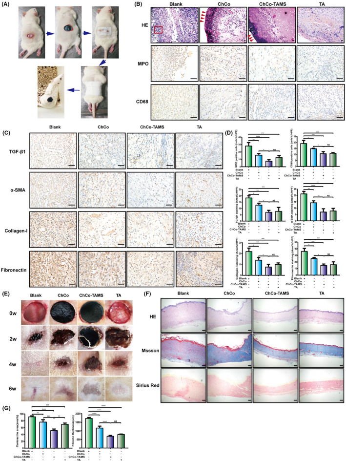
The anti‐fibrotic efficiency of ChCo‐based scaffolds in the rat dermal defect model (A) The procedures of dermal resection and scaffolds fixation on rats. (B) At the 7th day, the wound tissues from different groups were subjected to H&E and IHC staining to evaluate the pathological changes and infiltration of inflammatory cells (MPO‐, CD68‐positive); the red arrows indicated the attached composites and the frame suggested inflammatory cells; scale bar, 50 μm. (C) IHC assays were conducted to identify fibrotic‐related proteins, including TGFβ1, α‐SMA, collagen‐I and fibronectin; scale bar, 50 μm. (D) The quantification of the IHC results. (E) The tissue healing process in different groups was observed at indicated time points. (F) At the 6th week, the scar tissues were collected and assessed by H&E, Masson's trichrome and Sirius Red staining; scale bar, 200 μm. (G) The scar contraction and fibrotic thickness at the 6th week were quantified. The representative statistical results were performed using one‐way ANOVA test and shown as means ± SEM from 3 independent experiments. *P < 0.05, **P < 0.01, ***P < 0.001, ****P < 0.0001
