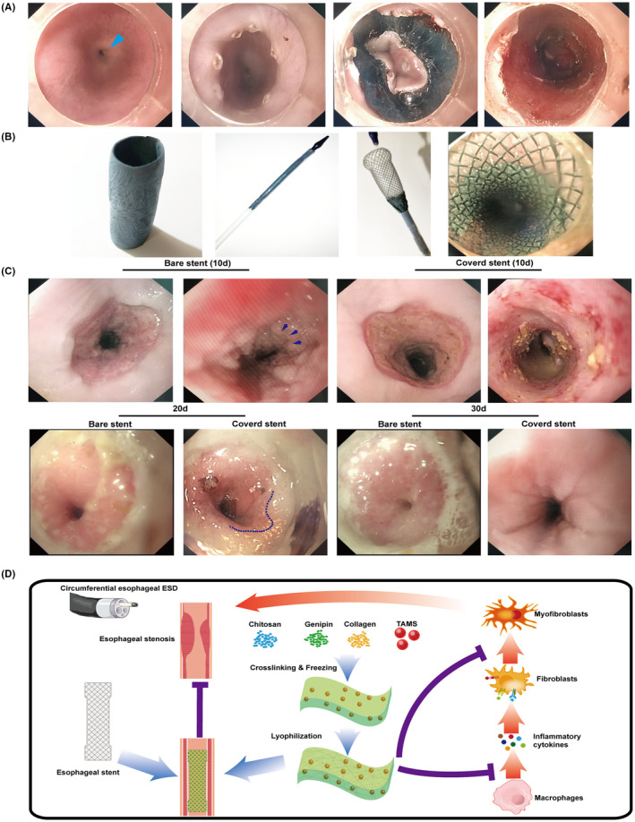
FIGURE 7.

Stricture ameliorating efficiency of ChCo‐TAMS scaffolds in the porcine oesophageal ESD model (A) The circumferential oesophageal ESD procedures. From left to right indicated: normal oesophageal lumen (Blue arrow indicated the cardia); marking of the cutting edge by electrocoagulation; circumferential cutting of the mucosal tissue at the oral side; the post‐operative wound bed. (B) Application of ChCo‐TAMS on post‐ESD wound. From left to right indicated: the prepared cylindrical ChCo‐TAMS; the folded oesophageal metal stent wrapped with ChCo‐TAMS; release of ChCo‐TAMS wrapped stent in vitro; attachment of ChCo‐TAMS onto the post‐ESD wound. (C) Observation of the wound healing with or without the application of ChCo‐TAMS at indicated time intervals. At the 10th day, the arrows indicated the newly regenerated epithelium; at the 20th day, the dotted line indicated the margin between normal mucosa and incompletely healed reddish mucosa. (D) The schematic diagram illustrated the manufacture and the potential mechanisms of the ChCo‐TAMS scaffolds in preventing ESD‐related oesophageal strictures
