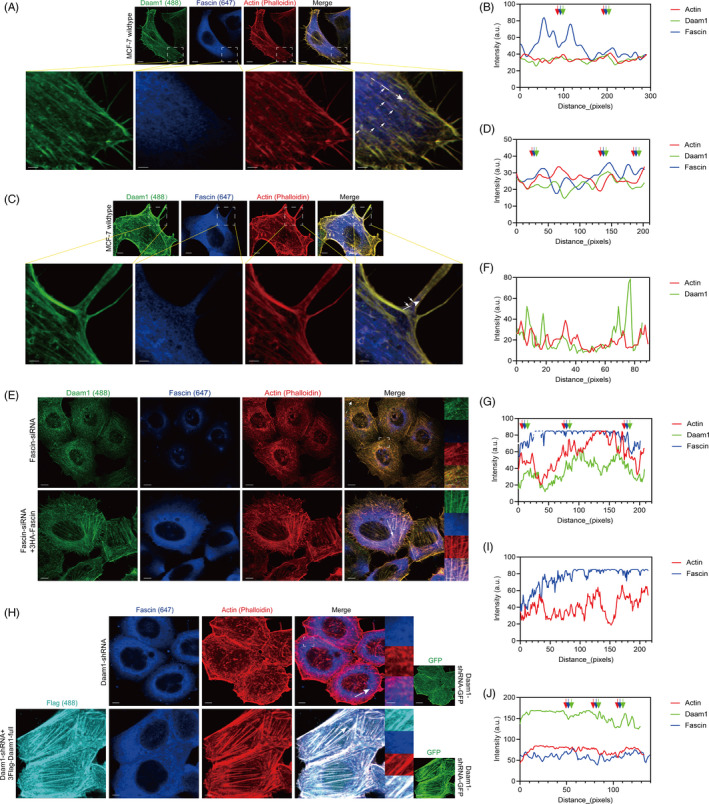
FIGURE 5.

Daam1 and Fascin recruit each other, and they cooperatively mediate actin filament assembly and pseudopodia formation. A‐D, Endogenous Daam1 (green) and Fascin (blue) in MCF‐7 cells were colocalized with actin filaments and pseudopodia (filopodia, red). Bar, 5 μm. The boxed region shows the enlargement of local actin filaments, and white arrowheads indicate colocalization. Bar, 100 pixel. Fluorescence intensity profiles of Daam1, Fascin and actin were measured at the positions marked by arrows in merged insets. E‐G, Fascin‐silenced MCF‐7 cells (Fascin‐siRNA) were immunostained with antibodies against Daam1 (green) and Fascin (blue) and compared to the rescue group (Fascin‐siRNA + 3HA‐Fascin). The actin filaments were stained with phalloidin (red). Bar, 5 μm. The boxed region showed the local enlargement state. Bar, 5 pixel. H‐J, Daam1‐silenced MCF‐7 cells (Daam1‐shRNA) were immunostained with antibodies against Daam1 (cyan) and Fascin (blue) compared to the rescue group (Daam1‐shRNA + 3FLAG‐Daam1‐full). ShRNA targeting Daam1 was labelled with GFP (green), and actin filaments were labelled with phalloidin (red). Bar, 5 μm. The boxed region showed the local enlargement state. Bar, 5 pixel
