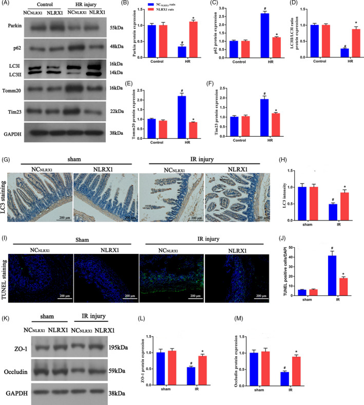
FIGURE 3.

NLRX1 promotes mitophagy and alleviates intestinal IR‐induced injury. A‐F, Western blotting experiments to assess mitophagy flux (parkin, p62 and LC3II/LC3I ratio) and mitochondrial protein (Tomm20 and Tim23) levels in intestinal tissues. G,H, Immunohistochemistry assay of LCⅡ in NCNLRX1 and NLRX1 overexpression rats. (scale bar = 200 μm; magnification ×100). I,J, Representative TUNEL staining and quantitative analysis after intestinal IR injury. Relative apoptotic rates are represented as TUNEL‐positive cells/DAPI (scale bar = 200 μm; magnification ×100). K‐M, Protein expression levels of ZO‐1 and occludin. #P < .05 vs sham group; *P < .05 vs NCNLRX1+IR group
