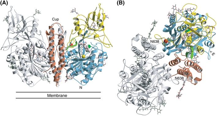
Figure 2.

A, Structure of GCPII (A, B). Three‐dimensional structure of the dimer. One subunit is shown in gray, while the other is colored according to organization into domains. Domain I (light blue), domain II (yellow), and domain III (brown). The dinuclear zinc cluster at the active site is indicated by dark green spheres, the Ca2+ ion near the monomer‐monomer interface by a red sphere, and the Cl− ion by a yellow sphere. The GPI‐18431 inhibitor is shown as small beige balls. The “glutarate sensor” (the β15/β16 hairpin) is shown in light green. The seven carbohydrate side chains located in the electron density maps are indicated. The position of the structure relative to the membrane is shown in (A). B, Provides a view into the “cup” of the dimeric enzyme. The entrance to the catalytic site is indicated (“E”). Reproduced with permission from Mesters et al34
