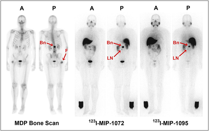
Figure 9.

Representative anterior A, and posterior (P) whole‐body planar images of patient with radiographically confirmed metastatic prostate cancer who received 740 MBq (20 mCi) of 99mTc‐methylene diphosphonate (MDP) (left), followed by 123I‐MIP‐1072 (middle) and 123I‐MIP‐1095 (right) administered at 370 MBq (10 mCi). Depicted are images acquired at 4 h after injection. Arrows indicate detection of confirmed lesions in bone (Bn) of lumbar spine and uptake in suggestive 7‐mm lymph node (LN). Image reference standard was placed next to right leg. Subject had previous hip replacement as demonstrated by uptake in head of right femur (F) only on bone scan. Reproduced with permission from Barrett et al97
