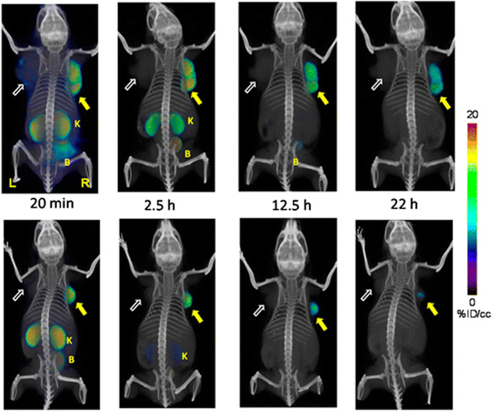
Figure 14.

Whole body PET‐CT imaging of PC3 PIP and PC3 flu tumor‐bearing mice with [64Cu]Lys‐NHCONH‐Glu inhibitor 6A as a building block (top row) and [64Cu]Lys‐NHCONH‐Glu inhibitor 6B (bottom row) at 20 min, 2.5 h, 12 h, and 22 h postinjection. Abdominal radioactivity is primarily due to uptake within kidneys and bladder. PIP = PC3 PSMA+ PIP (solid arrow); flu = PC3 PSMA− flu (unfilled arrow); K = kidney; L = left; R = right, B = bladder. All images are decay‐corrected and adjusted to the same maximum value. Reproduced with permission from Banerjee et al107
