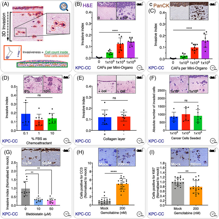
Figure 3.

Cancer cell invasion assay. A, Schematic overview of QuPath‐based scoring system. The number of cells which have invaded into the gel is calculated as an index of the total number of cells seeded to the top of the gel. B, Increasing the number of CAFs seeded to the Mini‐Organo during the contraction phase leads to an increase in the invasion of KPC cancer cells into the Mini‐Organo at the same timepoint. C, In addition to simple haematoxylin and eosin staining (see panel B), it is possible to use immunohistochemical staining for markers of interest such as for pan‐cytokeratin (which specifically stains the epithelial tumour cells) with data recapitulating that seen in H&Es. D, The effect of chemoattractants (such as FBS) added to the media can be determined using the Mini‐Organo. E, Some (not all) cancer cell lines will invade more readily to a thin layer of 1.5 mg/mL collagen I overlaid on top following transfer to the grids (+ col vs − col). KPC‐CCs invade regardless of the presence of this additional top layer. F, Increasing the number of cancer cells seeded on top of the plug does not increase the absolute number of cancer cells invaded into the plug; however, it will decrease the invasive index accordingly (see formula in Figure 3A). G, Therapeutic targeting of cancer cell invasion can also be tested in Mini‐Organos. Treatment of KPC‐CCs with the non‐muscle Myosin II inhibitor Blebbistatin during the invasion stage (drug not present during contraction phase) and shows significant decreases in cancer cell ability to invade into the remodelled Mini‐Organos. H, Evaluation of the effects of anticancer therapies on invaded cancer cells can also be tested. Treatment of KPC‐CC with standard‐of‐care gemcitabine leads to an increase in Cleaved Caspase3 (CC3)(Apoptosis). I, decrease in Ki67 (proliferation) as measured by IHC staining
