Figure 5.

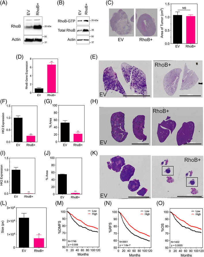
RhoB overexpression does not affect tumor growth in vivo but does contribute to a decreased metastasis. A. Immunoblot assays were performed using lysates prepared from MDA‐MB‐231 EV (empty vector) and a RhoB+ overexpressing subclone under 20% O2. B, RhoB‐GTP pulldown assay was performed to detect levels of activated RhoB in MDA‐MB‐231 EV and RhoB+ subclones under 20% O2. The amount of activated or total RhoB was detected by immunoblotting the pulldown samples and the whole cell lysates with a RhoB antibody. C, Tumor sections for the indicated subclones of MDA‐MB‐231 cells that were injected into the mammary fat pad of NSG mice were stained with hematoxylin in order to measure their area using ImageJ software (scale bar = 10 000 μm). D, Human RhoB content in the tumors was quantified by quantitative polymerase chain reaction. E, H, K. E, Whole‐mount inflated lungs (scale bar = 10 000 μm), (H) livers, and (K) lymph nodes were stained with hematoxylin and eosin to identify metastatic foci. Human genomic DNA content in the (F) lungs and (I) liver was quantified by quantitative polymerase chain reaction for human HK2 gene sequences. The percent area stained for hematoxylin for each (G) lung section and (J) liver section was measured using ImageJ software. L, The size or perimeter of each lymph node section was measured using ImageJ software. All data are shown as mean ± standard error of mean; n = 8. **, P < 0.01; ***, P < 0.001 vs. EV (student t test). Kaplan‐Meier analysis of (M) distant‐free metastasis (n = 1746), (N) relapse‐free survival (n = 3951), and (O) overall survival (n = 1402) of breast cancer patients stratified by RhoB mRNA expression above (high) or below (low) the median level is presented
