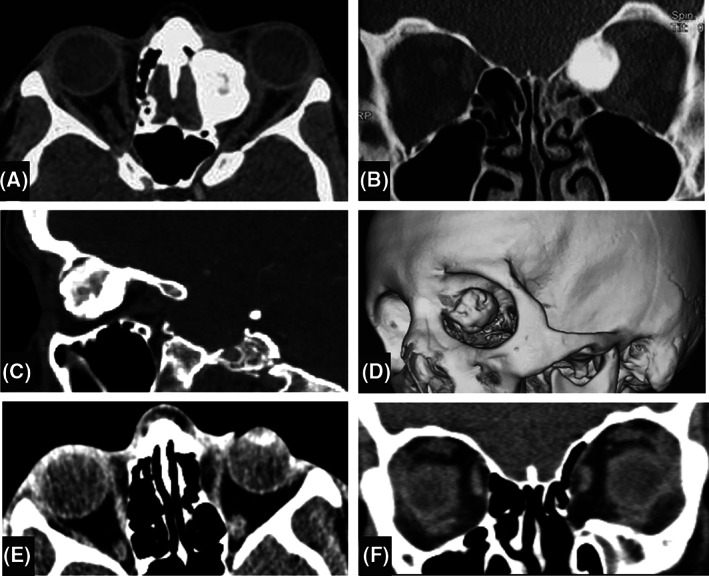
FIGURE 2.

Orbital CT scan of the patient. At presentation, a superonasal giant osteoma is seen at the junction of the left frontal bone and the ethmoidal sinus, extending to the extraconal space of the left orbit. Frontal sinuses are not pneumatized and left ethmoidal sinus is opaque and partially obliterated. Compressive effect of the mass is seen on the optic nerve and orbital soft tissues. A, axial view; B, coronal view; C, sagittal view; D, three dimensional oblique view. At final follow up, the orbital walls are intact excepting the lamina papyracea and the soft tissues have returned to normal position E and F
