FIGURE 3.

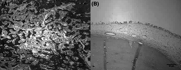
Illustrated microphotographs of the tumor showing dense mature predominately lamellar bone with peripherally located osteoblasts and inter lamellar fibro‐vascular tissue. A and B, hematoxylin and eosin

Illustrated microphotographs of the tumor showing dense mature predominately lamellar bone with peripherally located osteoblasts and inter lamellar fibro‐vascular tissue. A and B, hematoxylin and eosin