
FIGURE 1.
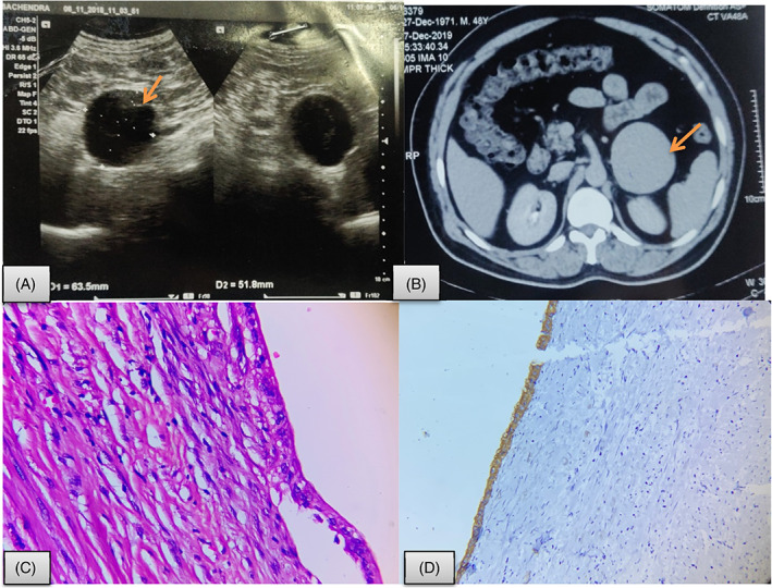
Epithelial adrenal cyst. A, USG abdomen showing (arrow) a well‐defined hypo‐echoic lesion in left suprarenal region measuring 64 × 52 mm (? adrenal origin); B, CECT showing a well defined, round to oval, hypo‐dense lesion with hyper dense thin wall measuring 6.3 × 6.4 × 6.6 cm in the retroperitoneum closely abutting pancreas (arrow); C, microphotograph displaying a cyst with fibro collage nous cyst wall lined by single layer of cuboidal epithelial cells (Hematoxylin and eosin; 40×); D, Immunohistochemistry shows CK positivity in lining epithelial cells (Hematoxylin and eosin; 400×)