
FIGURE 2.
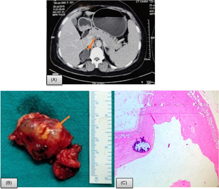
Adrenal pseudo cyst. A, CECT showing a multiloculated cystic lesion measuring 31 × 32 × 36 mm with multiple wall and septal calcification in right adrenal region (arrow); B, Resected specimen showing multiloculated lesion filled with fluid; C, Histopathology shows fibrocollagenous cyst wall with focal calcification and no lining. Remnant of adrenal tissue is seen at the outer aspect (Hematoxylin and eosin; 400×)