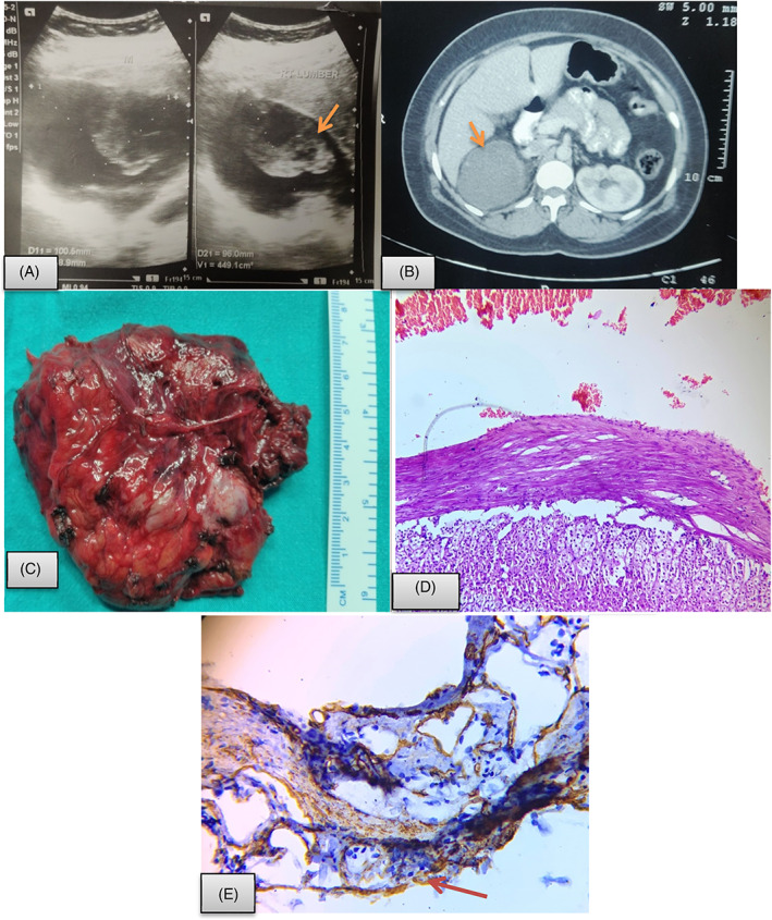
FIGURE 3.

Adrenal endothelial cyst. A, USG abdomen showing a mixed echogenic mass in right suprarenal region (arrow); B, CECT abdomen showed well‐defined right adrenal cyst as rounded hypo dense lesion measuring approx. 79 × 75 × 78 mm with internal haemorrhage as slight hyper dense echogenic area in it (arrow); C, Resected specimen showing rright suprarenal mass measuring 8 × 7 cm with cystic and solid component; D, Histopathological evaluation display cystic structure with fibrocollagenous wall lined by flattened endothelial cells and red blood cells in the lumen with adrenal tissue at the periphery (Hematoxylin and eosin; 40×). E, Immunohistochemistry show CD34 positivity in lining endothelial cells as well as in microvasculature of adrenal tissue (40×)
