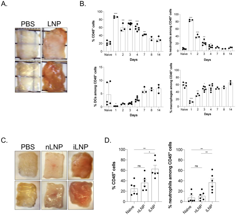
Figure 1. Intradermal inoculation with LNPs induces robust inflammation.
A. Intradermal inoculation with LNP induced visible levels of inflammation. Pictures were taken 24 hours post PBS or LNP injection. B. Skin samples from the mice injected with PBS or LNP were harvested at the indicated time points, analyzed by flow cytometry, and displayed as cell percentages. C. As in A, but LNPs with (iLNP) or without (nLNP) ionizable lipids were used. Unlike iLNPs the nLNPs induced no visible signs of inflammation. D. Skin samples from C were analyzed for leukocytic infiltration 24 hours post inoculation. For all the charts the data were pooled from two separate experiments and displayed as percent ± SD. Each dot represents a separate animal. Student’s two-tailed t-test was used to determine the significance between naïve and the experimental samples. ****p<0.0001, ***p<0.0005, **p<0.005, *p<0.05, ns = not significant. No differences were observed between samples harvested from naïve or PBS treated animals and are used interchangeably throughout the manuscript.

