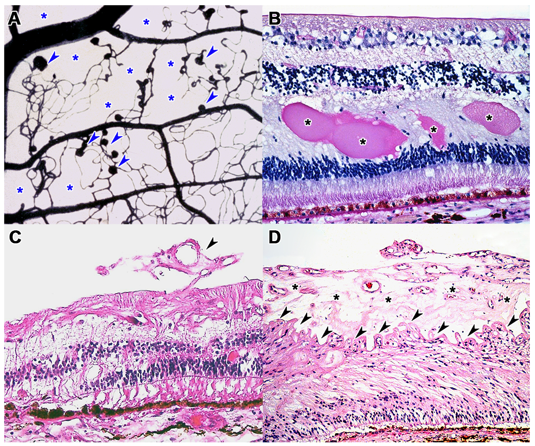
Figure 1: Histopathologic features of diabetic retinopathy.

(A) Trypsin digest from a human donor, stained with India ink, showing numerous microaneurysms (arrowheads) and large areas of capillary dropout (asterisks). (B) Hematoxylin and eosin stain showing lipoproteinaceous exudates in the outer plexiform layer (asterisks) in a patient with diabetic macular edema. (C) Early neovascular tuft (arrowheads) in an eye with early proliferative diabetic retinopathy. (D) Thick preretinal neovascular sheet (asterisks) with wrinkling of the ILM (arrowheads) in a patient with advanced proliferative diabetic retinopathy.
