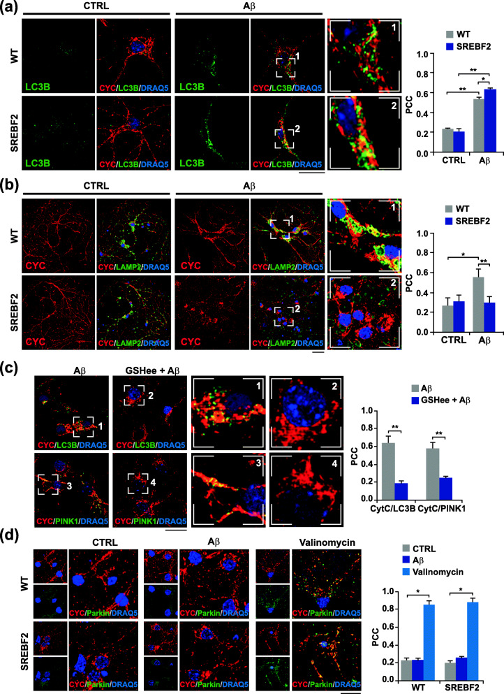
Fig. 2.
Cholesterol-induced depletion of mitochondrial GSH stimulates incomplete PINK1-mediated mitophagy in cultured primary neurons exposed to Aβ. Embryonic cortical and hippocampal neurons isolated from WT and SREBF2 mice were treated with Aβ (5 μM) for 24 h or valinomycin (10 μM) for 3 h to trigger mitophagy. a and b Representative confocal images of neuronal-enriched cultures double immunostained with antibodies for LC3B (green) and CYC (red) (a) and for LAMP2 (green) and CYC (red) (b). Scale bar: 25 μm. c SREBF2 cells were pre-incubated with GSH ethyl ester (GSHee, 0.5 mM) for 30 min prior mitophagy induction with Aβ. Shown are representative confocal images of double immunostainings for LC3B (green) and CYC (red) and for PINK1(green) and CYC (red). Scale bar: 25 μm. d Representative confocal images of a double immunolabeling for CYC (red) and parkin (green). Scale bar: 25 μm. Nuclei were counterstained with DRAQ5 (blue). Insets show a 3-fold magnification of the indicated regions. In all the cases, the Pearson’s correlation coefficient (PCC) was calculated from 3 independent experiments (at least 6 random fields were analyzed per condition). *P < 0.05; **P < 0.01 (data are mean ± SD)

