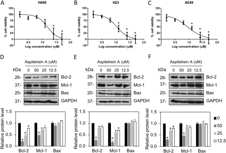
Fig. 2.
The effects of AA on the viability of human lung cancer cells. H460 (a), H23 (b), and A549 (c) cells were treated with AA (0, 1.5625, 3.125, 6.25, 12.5, 25.0 and 50 μM) for 24 h. Cell survival was evaluated by MTT assay. The plots present the percentage of cell viability ± SEM from three independent experiments (n = 3). *, p < 0.05 vs nontreated cells. H460 (d), H23 (e) and A549 (f) cells were treated with AA (0–50 μM) for 24 h. Anti-apoptotic Bcl-2 and Mcl-1 and pro-apoptotic Bax were examined by Western blot assay. Blots were reprobed with anti-GAPDH antibodies to confirm equal loading. The protein levels were quantified and normalized to the glyceraldehyde-3-phosphate dehydrogenase (GAPDH) level. The data are the mean ± SEM from three independent experiments (n = 3). *, p < 0.05 vs nontreated cells

