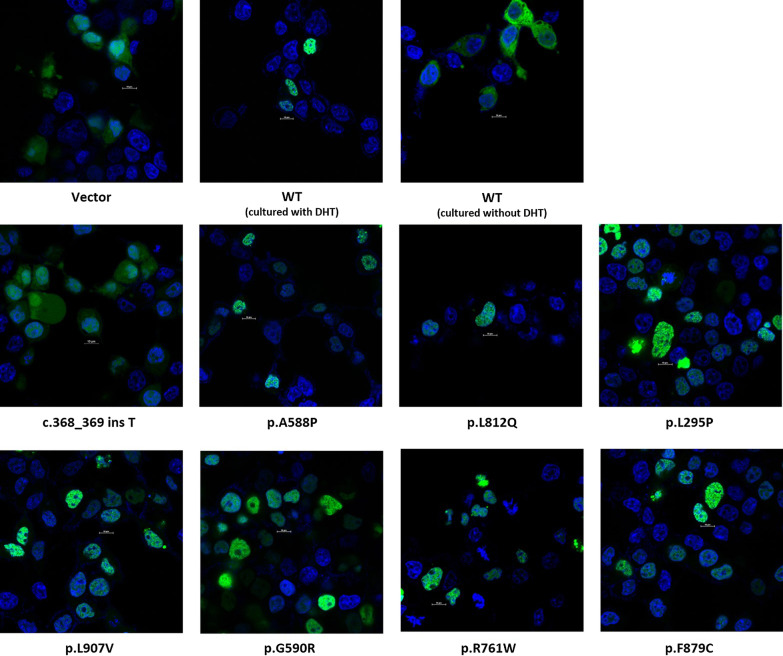
Fig. 4.
Nuclear translocation of GFP-tagged wild-type and mutant ARs. The upper panel from left to right: Empty vector and GFP-tagged WT AR (cultured without DHT) exhibited diffused localization in the cytoplasm. GFP-tagged WT AR (cultured with DHT) exhibited strong nuclear localization. The middle panel and lower pannel: Nuclear translocation of GFP-tagged eight mutant ARs in DHT treated cells. A similar nuclear localization pattern was observed in 7 novel missense mutants, while, the c.368_369 ins T mutant exhibited nuclear localization similar to empty vector. The scale bar represents 10 μm

