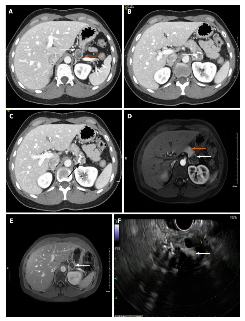
Figure 1.
Complex, cystic and solid mass in the pancreatic tail. A: Axial computed tomography with IV contrast showing a bilobed hypodense lesion with superior and inferior components with peripheral thick rim of enhancement. White arrow: Showing solid component. Orange arrow: Inferior cystic component; B: White arrow: Calcification within tumor; C: Black arrow: Compression of splenic vein by mass effect from tumor; D: Arterial phase magnetic resonance imaging (MRI) image. White arrow: Showing the tumor. Orange arrow: Normal pancreatic tissue; E: MRI post gadolinium study, white arrow: Thick peripheral solid enhancing component with central non-enhancing cystic component with internal septation; F: Solid and cystic mass on endoscopic ultrasound.

