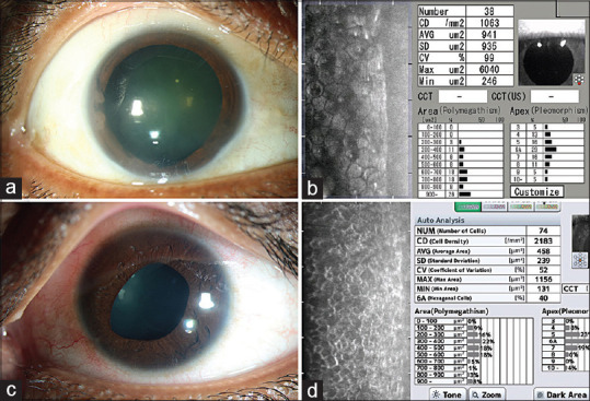
Figure 6.

Specular microscopy in ICE syndrome: (a and b) Slit lamp photograph (a) of a 45 year old patient with ICE syndrome showing broad peripheral synechiae, central to paracentral corneal haze in the inferotemporal quadrant; the specular image (b) from the superior mid peripheral cornea (clear area of the cornea) shows enlarged endothelial cells, rounding of the cellular boundaries and increased black out areas within the cells. (c and d) Slit lamp photograph (c) of a 25 year old patient with ICE syndrome; the specular microscopy (d) shows the characteristic dark-light reversal pattern
