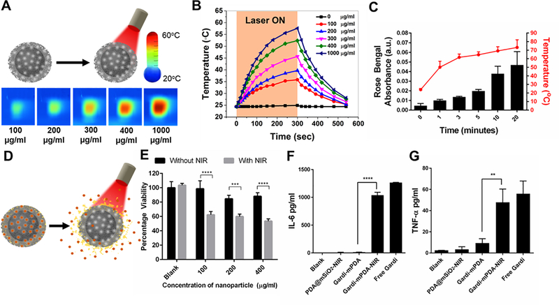
Figure 3.
(A) Schematic representation of NIR irradiation of pristine PDA@mSiO2 nanoparticles and IR images of temperature rise with increase in particle concentration after 5 minutes of NIR laser treatment. (B) Temperature profile and effect of PDA@mSiO2 particle concentration on temperature rise when aqueous solutions were subjected to laser power density of 14 mW/mm2. (C) Cumulative release of model dye from the PDA@mSiO2 nanoparticles after different laser irradiation durations and their corresponding solution temperature (laser power density, 14 mW/mm2). (D) Schematic representation of gardiquimod loaded PDA@mSiO2 (gardi-mPDA) nanoparticles and release of cargo with NIR treatment. (E) Cancer cell viability after treatment with gardi-mPDA with and without NIR. BMDC activation indicated by cytokine secretion (F) IL-6 and (G) TNFα. Data represented as mean ± SD. ** p<0.01, *** p<0.001 and **** p<0.0001 by one-way ANOVA with Tukey’s posttest.

