Figure 4.
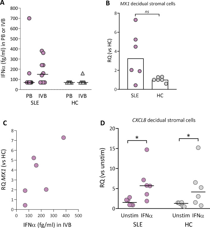
IFNα protein levels in peripheral or intervillous blood and MX1 gene expression in decidual stromal cells from women with SLE and healthy controls. (A) IFNα protein levels in plasma obtained from peripheral or intervillous blood at delivery in women with SLE and healthy controls (HCs). (B) Quantitative PCR analysis of MX1 gene expression in decidual stromal cells from women with SLE and HC. (C) Correlation between IFNα protein levels in plasma from intervillous blood and MX1 gene expression in decidual stromal cells from women with SLE. (D) Quantitative PCR analysis of CXCL8 gene expression in decidual stromal cells from women with SLE and HC stimulated with IFNα (1 ng/mL) or not. Relative quantification (RQ) was calculated using the average CXCL8 expression in unstimulated HC controls as a reference. Horizontal bars in A and D indicate medians, scatter plot bars in B display median and each symbol represents one individual. (B) Mann-Whitney U test and (D) *p≤0.05 Wilcoxon matched-pairs signed rank test.

