Fig. 1.

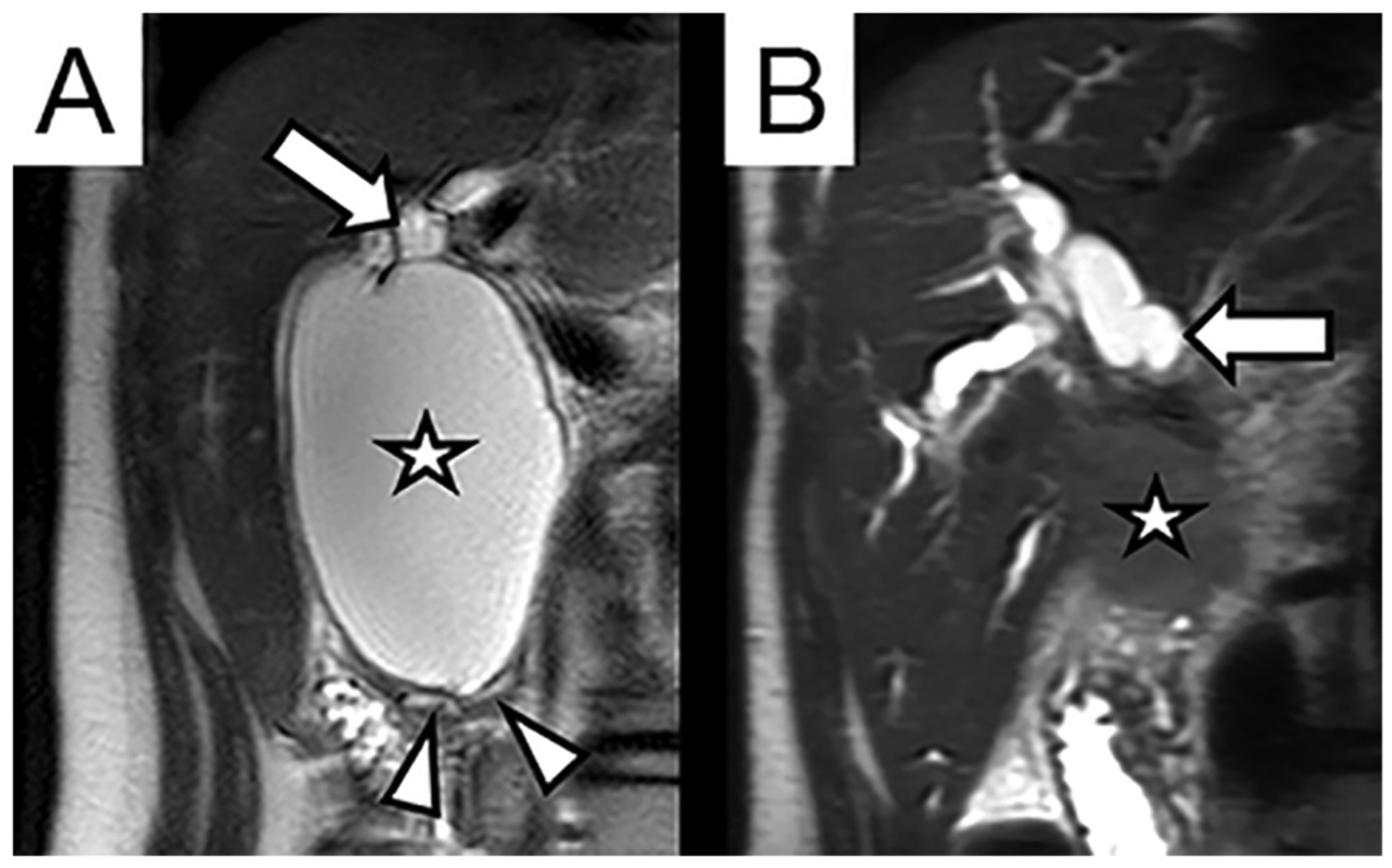
Coronal T2-weighted MRI images of the right upper abdomen. (A) 16-year-old female with two months of intermittent severe abdominal pain, found to have focal cystic dilation of the common bile duct (Type I choledochal cyst, annotated with star). The lumen of the cyst communicates with the dilated common hepatic duct (arrow). The cyst involves the distal intrapancreatic bile duct, resulting in splaying and displacement of the pancreatic parenchyma and pancreatic duct (arrow heads). (B) Same patient two years after cyst resection, presenting with new progressive pain, jaundice, and elevated serum bilirubin and CA19–9. MRI demonstrates new ill-defined infiltrative mass in porta hepatis (star) with severe intrahepatic biliary dilation (arrow).
