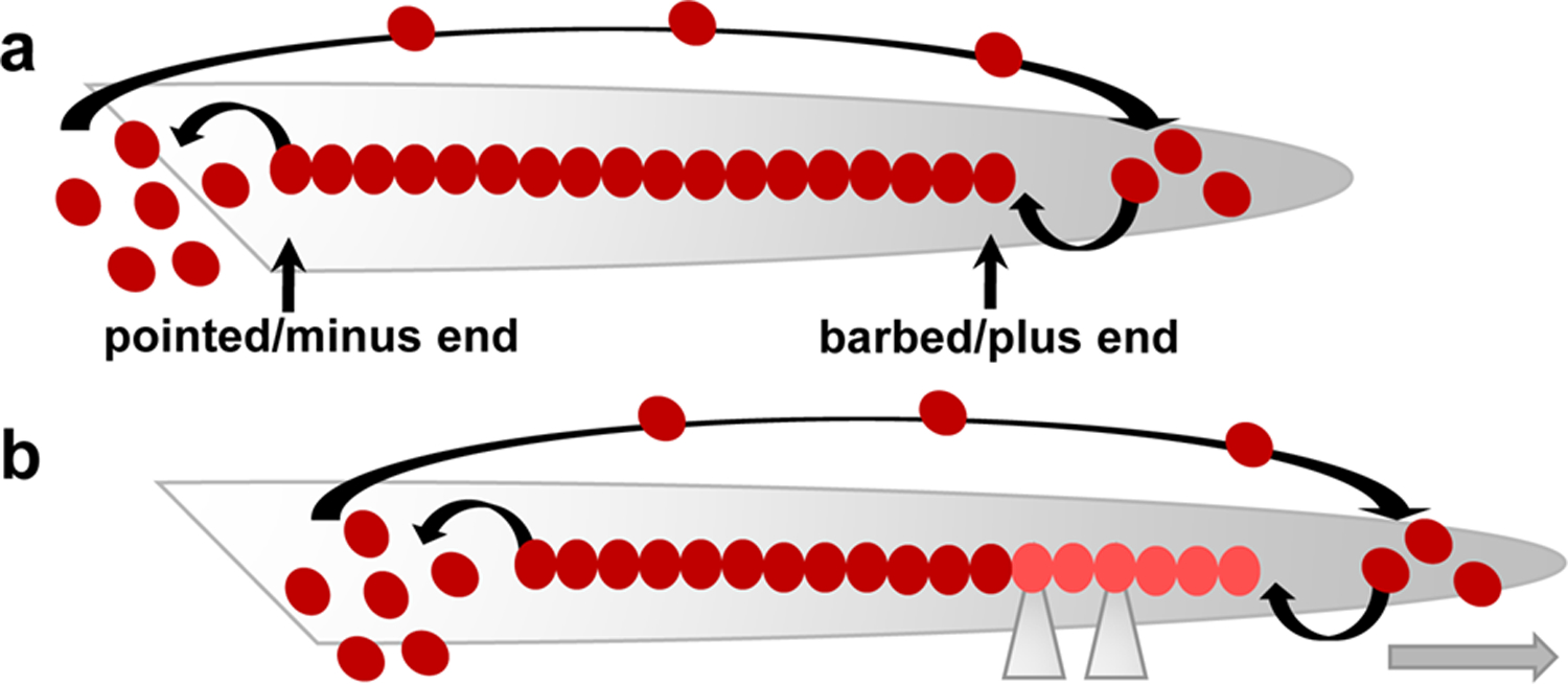
Figure 1. Diagram illustrating actin dynamics during treadmilling and forward protrusion of cellular processes.

(a) F-actin filament growth occurs at the barbed/plus end and is balanced by net filament disassembly at the pointed/minus end and by recycling of G-actin subunits back to the barbed/plus end (large arrow at the top). Thus, F-actin filaments coexist with G-actin monomers at a ‘steady state’. (b) When the actin filament is anchored to a growth promoting (permissive) substrate via the action of adhesion complexes (grey triangles), actin retrograde flow (from the barbed/plus end to the pointed/minus end; not shown) is reduced, and the F-actin filament seemingly moves in one direction, leading to forward movement of cellular protrusions (grey arrow). Actin monomers are depicted as dark red filled circles; newly polymerized F-actin is shown in light red
